On 23 February 2026, the Equality High Court in Cape Town granted Doctors for Life International (DFL) permission to participate as amicus curiae (a friend of the court) in a case concerning alleged “transgender” discrimination at a high school in the Western Cape. The Court is allowing DFL to present expert medical evidence showing that gender is rooted in, and inseparable from, biological sex. The evidence will also show that the practice of socially transitioning children to a gender opposite to their biological sex as a treatment for gender dysphoria is harmful and has been discredited in the international medical community.
The experts will refer to recent international developments, including the 2024 Cass Review in the United Kingdom, which found that the scientific evidence supporting gender-transition interventions for minors is weak and uncertain. There has also been growing controversy surrounding the WPATH Standards of Care following what has become known as the “WPATH Files”. In addition, several European countries have recently limited gender-transition interventions for children due to the harm it can do.
The case is scheduled to be heard in the Cape Town High Court in October 2026.
The following story recounts the lives of the four Mkhize (surname) children at the Table Mountain Orphan Centre close to Pietermaritzburg in KwaZulu-Natal Midlands. The day care centre is part of Doctors For Life orphan program called LifeChild.
They were born in the Table Mountain village and raised by their mother within a large extended family of approximately 20 members, including their grandparents. Over time, the family experienced repeated and profound tragedies, including the deaths of multiple relatives due to illnesses and motor vehicle accidents, in some instances losing two or three family members on the same day. These sudden losses left the family deeply traumatized, and eventually only three adults and the four children remained.
Although their mother was initially alive, she later remarried and left the children in the care of their grandmother. Sadly both the grandmother and the mother later passed away, leaving the children under the responsibility of their aunt, who was only 16 years old at the time. She was compelled to discontinue her education to care for the children. The household faced extreme deprivation, including food shortages, inadequate clothing, and the inability to access government grants due to the absence of identity documentation of their mother. As a result, the children were unable to attend school due to lack of uniforms and school expenses. At the age of 7 years old, the eldest child began herding cattle for others to earn income to provide food for his siblings, while the younger children were depended on neighbours for meals.
During this desperate time, God led the Doctors For Life organization to establish a centre in the Table Mountain area. The four children were taken in by LifeChild and were provided with consistent nutrition and access to education. Many other children being taken care of by Doctors For Life have similarly painful backgrounds of how they came to the organization. Through the dedicated work of Doctors For Life, this centre not only provides essential physical care but also spiritual support enabling them to grow, heal, and live happily.
We request that you continue praying for the Lord’s work at the Table Mountain centre-that He may sustain, guide, and bless this ministry so that it may continue to serve vulnerable children and transform lives for His glory. Thank you for your continued support and prayers. Written by Nelisiwe Hadebe, caretaker at the Table Mountain centre)
Flooding in southern Mozambique continues to place immense pressure on already vulnerable communities, particularly around Xai-Xai and the Limpopo River basin. Government hospitals in the area remain affected by ongoing nursing strikes, which has led to a significant increase in patients seeking care at our clinic. While the clinic itself has not been directly flooded, access remains difficult due to severe damage to surrounding road infrastructure. Many of the main asphalt roads are badly degraded, with large potholes slowing travel and limiting reliable access. The situation on major transport routes remains critical. The EN1 highway – Mozambique’s main north-south artery – is still cut near Xai-Xai. Repair work close to the Nguluzane River bridge has been repeatedly delayed due to strong water currents. An alternative inland route briefly reopened earlier this month, but sections of it failed again on 5 February, leaving hundreds of vehicles stranded and severely disrupting the delivery of food, medical supplies, fuel, and other essential goods. Fuel shortages are now widespread in the region. The broader humanitarian impact is severe. Many families have lost their homes, crops, and remaining food supplies, increasing hunger and vulnerability. The risk of cholera and other water-borne diseases is extremely high, particularly in areas with unsafe drinking water and limited sanitation. Until recently, some communities were still completely isolated and only reachable through rescue operations. National figures indicate that approximately 185 people have lost their lives as a result of flooding and related disease outbreaks since October. An estimated 845,000 people have been affected across the country, with tens of thousands of homes damaged or destroyed and extensive damage to roads and critical infrastructure. There are, however, cautious signs of improvement. Water levels in the Limpopo River have begun to fall, and assessments are underway on the elevated road between Chimcumbana and the city of Xai-Xai. While not yet confirmed, there are indications that some damaged road sections may reopen soon, which would significantly improve access for relief efforts and supply deliveries. We continue to monitor the situation closely and adjust our response as conditions evolve. We are deeply grateful for the ongoing support shown by our community during this challenging time.
Johann
(Aid to Africa) Doctors For Life International P.O. Box 6613 Zimbali 4418 South Africa +27 834582949 (mobile) +27 (0) 32 481 5550/1/2/3 (office) +27 (0) 32 481 5554 (fax) www.doctorsforlife.co.za<http://www.doctorsforlife.co.za> Www.dfl.org.za<http://Www.dfl.org.za>
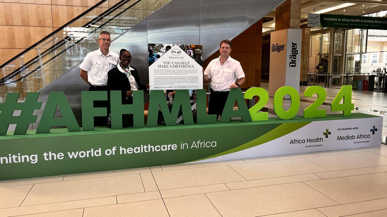
DFL was invited to present its stance on abortion at the recent Africa Health Conference and Medlab, held at the ICC in Cape Town. In addition, DFL was provided with a complimentary exhibitor's table to showcase its work.
Nearly 500 exhibitors of medical products from over 50 nations were expected at this international conference, with more than 7,000 visitors anticipated over the course of three days, including approximately 3,000 delegates. DFL had the opportunity to engage with many exhibitors and attendees.
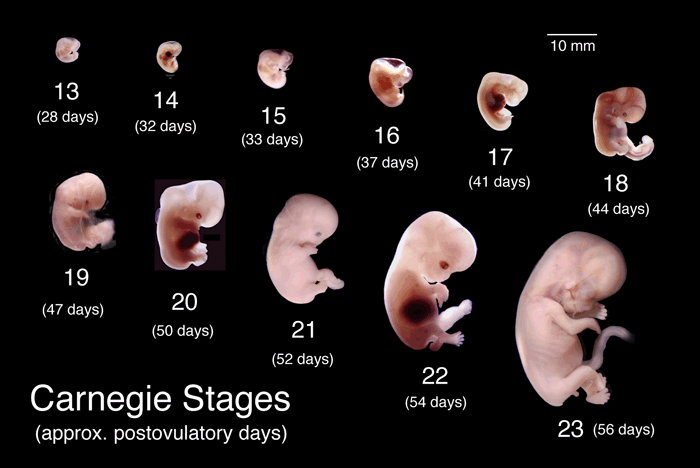
Dr. Monica Molatlegi represented DFL during the ethical session and participated in the debate on abortion. She laid the foundation for the pro-life stance by explaining that life begins at fertilization, referencing the Carnegie stages of embryonic development, which is considered the gold standard in embryology. Her insights received enthusiastic support from several delegates.
The Africa Health Conference also invited DFL to participate in a debate and open Q&A session on abortion during one of the ethical panels. Dr. Monica Molatlegi represented DFL, effectively presented the organization’s stance on abortion.
Here are additional photos of the other exhibitors at the conference.
Johan Claassen and Francois Williams were manning the DFL exhibit table and engaging with visitors.
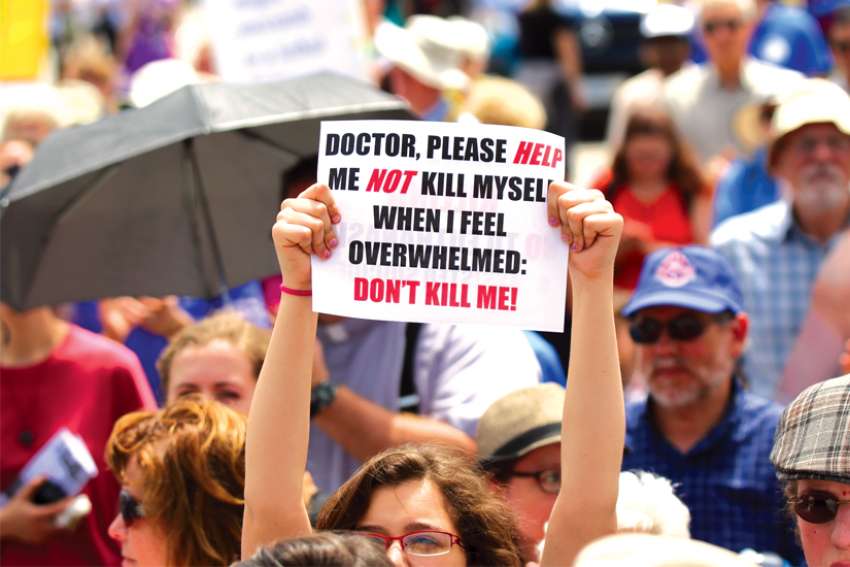
The term “assist” inherently implies providing support or aid in an action. In contrast, “suicide” specifically refers to the deliberate and voluntary act of ending one’s life, focusing on the action itself, not the motivation behind it. Therefore, the term “assisted suicide” is both accurate and descriptive. The key point remains suicide is suicide, and its outcome remains the same—life ceases. Terminology is manipulated to obscure the nature of euthanasia and assisted suicide, emphasizing the use of the euphemism “medical aid in dying” (MAID). The terminology shift is designed to legitimize the act and reduce stigma by associating it with medicine. The true intention behind these linguistic changes is to encourage more assisted suicides.
The EU’s executive arm unveiled a plan to require online platforms to detect and report the sharing of child sex abuse images on the internet. This would force companies operating in the EU to detect, report and remove the material. An increase in online child sex abuse has been noticed globally. Reports of internet child abuse rose from 1 million to almost 22 million during 2014-2020 and over 65 million images and videos of child sexually abused images were identified. Detection, reporting and removal of child sexual abuse online is also urgently needed to prevent the sharing of images and videos of the sexual abuse of children, which retraumatizes the victims often years after the sexual abuse has ended.”
In response to the Canadian government’s recent decision to permit Medical Aid in Dying (MAiD) for people with mental illnesses, the Canadian Mental Health Association (CMHA) issued a statement expressing deep disappointment. According to CMHA, until the health care system adequately responds to the mental health needs of Canadians, assisted dying should not be an option. This they also testified before the Senate in November of 2020.
CMHA points out that it is “not possible to determine whether any particular case of mental illness represents “an advanced state of decline in capabilities that cannot be reversed.” And the CMHA “know that cases of severe and persistent mental illness that are initially resistant to treatment can, in fact, show significant recovery over time. Mental illness is very often episodic. Death, on the other hand, is not reversible. In Dutch and Belgian studies, a high proportion of people who were seeking MAID for psychiatric reasons, but did not get it, later changed their minds.”
United Nations Human Rights experts expressed alarm at a growing trend to legalise euthanasia based largely on having a disability or disabling conditions, including in old age. They said that Disability should never be a ground or justification to end someone’s life directly or indirectly.
This, the experts said, that normalizing euthanasia for the people who are not terminally ill or suffering at the end of their lives would give rise to discriminative assumptions about the inherent “quality of life” or “worth” of the life of a person with a disability. Such assumptions are grounded in ableism and associated stereotypes. Disability is not a burden or a deficit of the person. It is a universal aspect of the human condition.
The known facts of the science of human embryology are not “new”. The first to study the human embryo systematically was Wilhelm His, Sr., who established the basis of reconstruction, i.e., the assembling of three-dimensional form from microscopic sections. His, who has been called the “Vesalium of human embryology,” published his three-volume masterpiece Anatomie menschlicher Embryonen in 1880-85 [His, Vogel, Leipzig]. In it the human embryo was studied as a whole for the first time internationally. A detailedHandbook of Human Embryology by Keibel and Mall appeared in 1910-12[1] . Franklin P. Mall, who studied under His, established the Carnegie Embryological Collection in Baltimore and was the first person to stage human embryos (in1914)
The standard source for generations for the documentation for both human sexual and the human asexual reproductive technique of “twinning” has been the Carnegie Stages,[1] and especially when they developed their internet online website, volumes of articles and books have been written referencing those Stages and the URLs where they could be found. Those online URLs have now changed, precluding others reading these articles and books from double-checking them for accuracy. In order to continue to be able to verify such documentation, the following NEW URLs for theCarnegie Stagesis provided:
— See Chart of all 23 Stagesof the early developing human embryo, at: http://www.medicalmuseum.mil/index.cfm?p=collections.hdac.anatomy.index. Click into the “textbook” at the bottom left side of the screen to access more extensive details of each stage and the extensive scientific references.
Go to the URLs listed below (examples of first 6 Stages only). Any particular Stage will first be shown as a brief summary of the scientific facts on that website URL. But if you look toward the bottom left of the webpage, you will see a “photo” of a human embryology textbook by O’Rahilly and Muller; it is actually a hyperlink. Click into that hyperlink, and you will be taken to the full original pages of the Carnegie Stage giving extensive details and documentation.
A. Examples of new online URLs for the Carnegie Stages
Stage 1 is the unicellular embryo that contains unique genetic material and is an individually specific cell that has the potential to develop into all of the subsequent stages of a human being. It is the beginning of embryonic life and ontogenetic development that starts when an oocyte, arrested in metaphase of meiosis II, is penetrated by a sperm.[1]This is the first event of fertilization. The embryo has a postovulatory age of approximately one day, is between 0.1 to 0.15 mm in diameter and weighs approximately 0.004 mg.
“… Stage 1 is divided into three substages; a, b and c. Stage 1a is referred to as the primordial embryo[5] since all the genetic material necessary for the new individual, plus some redundant chromosomes, is now within a single plasma lemma (cell membrane). [Note:all of the components define the “embryo”, not just the genes; and these components must work in sync with each other, thus themselves pre-determining the final coding of the genome – which itself can change from internal and external causes during early development. The human genome is defined as including all the DNA in a cell — both nuclear and mitochondrial — not just the nuclear.][6] From the perspective of the female gamete it has also been named the penetrated oocyte. The fertilizing sperm has passed through the zona (capsula) pellucida and its plasmalemma has fused with that of the oocyte.
“… Penetration activates the embryo into resuming its arrested meiosis II [7] and after anaphase it enters telophase with the expulsion of the redundant chromosomes as a second polar body. This marks the beginning of Stage 1b in which the single-cell is referred to as the pronuclear embryo[8] . From the perspective of the female gamete it has also been named the ootid because its female component is haploid like a spermatid. However, in the pronuclear embryo there are two separate haploid components: one maternal, or female, pronucleus and one paternal, or male, pronucleus.
“… The pronuclei move toward each other and eventually compress their envelopes where they lie adjacent near the center of the cell. Stage 1c is the last phase of fertilization and exists for a relatively short period[9]. The pronuclear envelopes disappear and the parental chromosomes that were contained in separate pronuclei come together in a process called syngamy thereby establishing the genome of the embryo. The one-cell Stage 1c embryo is named the syngamic embryo orzygote. The chromosomes assume positions on the rapidly formed first mitotic spindle in preparation for cleavage.”
II. Additional Resources
A. Online URLs for the International Committee on Human Embryology
The most recent updating of theCarnegie Stages(Jan. 2012) by the international nomenclature committee on human embryology, i.e., the Terminologia Embryologica Committee (FIPAT)[1] , is now also online and accessible on the internet (although not as “user friendly”).
The embryo sexually reproduced begins to exist at the beginning of the process of fertilization:
To use this new website for the Terminologia Embryologica online go to FIPAT, at: http://www.unifr.ch/ifaa/. Click on “Free access to published terminologies”, “Enter” to get to: http://www.unifr.ch/ifaa/Public/EntryPage/HomePublic.html. You are now on the Public Entry Page; Click into “Source terminologies as originally published”, to get to: http://www.unifr.ch/ifaa/Public/EntryPage/ViewSource.html. This page lists the 3 Terminologias. To the right of the page, under “Terminologia Embryologica, from internal document (2009)”, click onto “General Terms”, that takes you to:http://www.unifr.ch/ifaa/Public/EntryPage/ViewTE/TEe01.html. At the bottom of the page see “Footnote #5: E1.0.1.0.0..0.2 – Aetas a fecundiatione– Fertilization age begins at the time of fertilization with the sperm penetrating the oocyte … . It is the age of the conceptus and the preferred measure.”
There is no such thing as a “pre-embryo”:
Again, go to FIPAT, at: http://www.unifr.ch/ifaa/. Click on “Free access to published terminologies”, “Enter” to get to: http://www.unifr.ch/ifaa/Public/EntryPage/HomePublic.html. You are now on the Public Entry Page; Click into “Source terminologies as originally published”, to get to: http://www.unifr.ch/ifaa/Public/EntryPage/ViewSource.html. This page lists the 3 Terminologias. To the right of the page, under “Terminologia Embryologica, from internal document (2009)”, click onto e2.0: “Ontogeny”in order to get to: http://www.unifr.ch/ifaa/Public/EntryPage/ViewTE/TEe02.html. You are now viewing “Page 8”. This is a bit tricky: Now use button-arrows at top right of web page to move to Page 10to arrive at the description of Carnegie Stages 1-5 in the Chart. The right side of the Chart provides the following documentation of the first 5 Stages; see especially “Single cellEMBRYO [St. 1] [[“Stage One”]]. At the bottom of Page 10, in a footnote, you can find their rejection of the false scientific term “pre-embryo”: Footnote #32 – E2.0.1.2.0.0.3 – Embryo praegastrulationis [St. 1 ad 6a] The term pregastrulation embryo is useful because such an embryo has distinctive attributes (see footnote 35). The foreshortened term “pre-embryo”, which has been used in legal and clinical contexts, is not recommended.”[[Note: “St. 1 ad 6a” means “Stages 1 to 6a“]]
was originally developed as a collaboration between embryologist Dr. Raymond Gasser at LSUHSC and the HDAC in Washington DC. The overall aim of the project is to make the Carnegie collection, which is housed at the HDAC, accessible for research and teaching of human embryology. Dr. John Cork at LSUHSC joined the project at its inception as the software developer with a special interest in 3D-reconstruction. The project has two components, both of which are supported by grants from the National Institutes of Health. Anyone can access the various stages of the new sexually reproduced developing human embryo by going to the Virtual Human Embryo’s “DREM DEMOS” page, click into “Enter”, then click into “Demo” on the left of the page. Click into “Stage One: Introduction”: https://www.prenatalorigins.org/virtual-human-embryo/stage.php?stage=1
Stage 1 is the unicellular embryothat contains unique genetic material and is an individually specific cell that has the potential to develop into all of the subsequent stages of a human being. It is the beginning of embryonic life and ontogenetic development that starts when an oocyte, arrested in metaphase of meiosis II, is penetrated by a sperm. This is the first event of fertilization.The embryo has a postovulatory age of approximately one day, is between 0.1 to 0.15 mm in diameter and weighs approximately 0.004 mg.
Fertilization is a series of events that begins when a sperm makes contact with an oocyte and ends with the intermingling of paternal (male) and maternal (female) chromosomes on the spindle at metaphase of the first mitotic division of the single cell. The events of fertilization require just over 24 hrs. to complete and normally take place in the ampulla of the uterine tube. Stage 1 is divided into three substages; a, b and c. Stage 1a is referred to as the ‘primordial embryo’since all the genetic material necessary for the new individual, plus some redundant chromosomes, is now within a single plasmalemma (cell membrane). From the perspective of the female gamete it has also been named the penetrated oocyte. The fertilizing sperm has passed through the zona (capsula) pellucida and its plasmalemma has fused with that of the oocyte. Penetration activates the embryo into resuming its arrested meiosis II and after anaphase it enters telophase with the expulsion of the redundant chromosomes as a second polar body. This marks the beginning of Stage 1b in which the single-cell is referred to as the ‘pronuclear embryo’. From the perspective of the female gamete it has also been named the ootid because its female component is haploid like a spermatid. However, in the pronuclear embryo there are two separate haploid components: one maternal, or female, pronucleus and one paternal, or male, pronucleus. The pronuclei move toward each other and eventually compress their envelopes where they lie adjacent near the center of the cell. Stage 1c is the last phase of fertilization and exists for a relatively short period.The pronuclear envelopes disappear and the parental chromosomes that were contained in separate pronuclei come together in a process called syngamy thereby establishing the genome of the embryo. The one-cell Stage 1c embryo is named the ‘syngamic embryo’ or zygote.The chromosomes assume positions on the rapidly formed first mitotic spindle in preparation for cleavage. [http://virtualhumanembryo.lsuhsc.edu/demos/Stage1/Intro_pg/Intro.htm]
C. The iPhone App – “Embryo”[*** in process of being changed to a new URL]
Thanks to USA.gov, anyone can access the new iPhone appentitled, “Embryo”, from the National Library of Medicine, documenting the same facts of human embryology. The iPhone app is now available at: http://apps.usa.gov/embryo/. It is necessary to have iTunes for this application, but you can download it free from this same site (also downloads onto all computers). As noted on this government website:
Scientists and educators have used the Carnegie Embryo Collection, housed at the National Museum of Health and Medicine, to define normal human embryo development for decades. A database, called the Virtual Human Embryo, has been created to provide digital serial sections of human embryos from the collection….
This project is a collaboration between:
National Library of Medicine
Eunice Kennedy Shriver National Institute of Child Health and Human Development
Louisiana State University Health Sciences Center
National Museum of Health & Medicine, Human Developmental Anatomy Center [TheCarnegie Stages of Early Human Embryonic Development]
III. A Few Additional References for Sexual and Asexual Human Reproduction:
Human Sexual Reproduction:
** Ronan O’Rahilly and Fabiola Muller, Human Embryology & Teratology, 3rd ed. (New York: Wiley-Liss, 2001):
— Recapitulation, the So-Called Biogenetic Law.The theory that successive stages of individual development (ontogeny) correspond with (“recapitulate”) successive adult ancestors in the line of evolutionary descent (phylogeny) became popular in the nineteenth century as the so-called biogenetic law. This theory of recapitulation, however, has had a “regrettable influence on the progress of embryology”(G. de Beer)…. According to the “laws” of von Baer, general characters (e.g., brain, notochord) appear in development earlier than special characters(e.g., limbs, hair). Furthermore, during its development an animal departs more and more from the form of other animals. Indeed, the early stages in the development of an animal are not like the adult stages of other forms but resemble only the early stages of those animals.The pharyngeal clefts of vertebrate embryos, for example, are neither gills nor slits. Although a fish elaborates this region into gill slits, in reptiles, birds, and mammals it is converted into such structures as the tonsils and the thymus (p. 16).
… (Fertilization is)the procession of events that begins when a spermatozoon makes contact with a secondary oocyte or its investments, and ends with the intermingling of maternal and paternal chromosomes at metaphase of the first mitotic division of the zygote. (p. 19).
— Although life is a continuous process, fertilization… is a critical landmark because, under ordinary circumstances, a new, genetically distinct human organism is formed… (p. 31).
— Fertilization takes place normallyin the ampulla (lateral end) of the uterine tube (p. 31).
— “The term ‘pre-embryo’ is not used herefor the following reasons: (1) it is ill-definedbecause it is said to end with the appearance of the primitive streak or to include neurulation; (2) it is inaccuratebecause purely embryonic cells can already be distinguished after a few days, as can also the embryonic (not pre-embryonic!) disc; (3) it is unjustifiedbecause the accepted meaning of the word embryo includes all of the first 8 weeks; (4) it is equivocalbecause it may convey the erroneous idea that a new human organism is formed at only some considerable time after fertilization; and (5) it was introduced in 1986 ‘largely for public policy reasons’(Biggers).”… Just as postnatal age begins at birth, prenatal age begins at fertilization,” (p. 88).
— “Undesirable terms in Human Embryology”: “Pre-embryo”; ill defined and inaccurate; Use “embryo”(p. 12). [Note: O’Rahilly is one of the originators of The Carnegie Stages of Early Human Embryological Development, and has sat on the international committee for human embryology for decades]
** BRUCE M. CARLSON, Human Embryology and Developmental Biology (St. Louis, MO: Mosby, 1994): Human pregnancy begins with the fusion of an egg and a sperm. (p. 3); … finally, the fertilized egg, now properly called an embryo, must make its way into the uterus (p. 3)[2] ; … The sex of the future embryo is determined by the chromosomal complement of the spermatozoon … Through the mingling of maternal and paternal chromosomes, the zygote is a genetically unique product of chromosomal reassortment .. (p. 31); … “After the eighth week of pregnancy the period of organogenesis (embryonic period) is largely completed and the fetal period begins. (p. 407)
** BRUCE M. CARLSON, Human Embryology & Developmental Biology (St. Louis, MO: Mosby, 1999): “Human pregnancy begins with the fusion of an egg and a sperm, but a great deal of preparation [recedes this event. First both male and female sex cells must pass through a long series of changes (gametogenesis) that convert them genetically and phenotypically into mature gametes, which are capable of participating in the process of fertilization. Next, the gametes must be released from the gonads and make their way to the upper part of the uterine tube, where fertilization normally takes place. … Finally, the fertilized egg, now properly called an embryo, must make its way into the uterus ….”. (p. 2); … Fertilization age: dates the age of the embryo from the time of fertilization. (p. 23) … In the female, sperm transport begins in the upper vagina and ends in the ampulla of the uterine tube [fallopian tube] where the spermatozoa make contact with the ovulated egg. (p. 27) … After the eighth week of pregnancy the period of organogenesis (embryonic period) is largely completed, and the fetal period begins.” (p. 447). … The sex of the future embryo is determined by the chromosomal complement of the spermatozoon. (If the sperm contains 22 autosomes and an X chromosome, the embryo will be a genetic female, and if it contains 22 autosomes and a Y chromosome, the embryo will be a male.) … Through the mingling of maternal and paternal chromosomes, the zygote is a genetically unique product of chromosomal reassortment, which is important for the viability of any species. (p. 32)
** WILLIAM J. LARSEN, Human Embryology (New York: Churchill Livingstone, 1997): In this text, we begin our description of the developing human with the formation and differentiation of the male and female sex cells or gametes, which will unite at fertilization to initiate the embryonic development of a new individual. … Fertilization takes place in the oviduct [not the uterus]… resulting in the formation of a zygote containing a single diploid nucleus. Embryonic development is considered to begin at this point. (p. 1); … “These pronuclei fuse with each other to produce the single, diploid, 2N nucleus of the fertilized zygote. This moment of zygote formation may be taken as the beginning or zero time point of embryonic development. (p. 17).
** Geoffrey Sher, Virginia Marriage Davis, Jean Stoess, In Vitro Fertilization:The A.R.T. of Making Babies (New York: Facts On File, 1998): The moment a sperm penetrates the egg’s zona pellucida, a reaction in the egg fuses the zona and the perivitelline membrane into an impermeable shield that prevents other sperm from entering. … Propelled by contractions of the fallopian tube, the dividing embryo begins its three- or four-day journey back to the uterus and continues to divide after it reaches the uterus. (The fertilization process occurs near the middle of the fallopian tube — not in the uterus.) (p. 18)
** Lewin, Benjamin, Genes VII(New York: Oxford University Press, 2000):
— “A genome consists of the entire set of chromosomes for any particular organism, and therefore comprises a series of DNA molecules, each of which contains a series of many genes. The ultimate definition of a genome is to determine the sequence of the DNA of each chromosome. (p. 4)
… “Genes not residing within the nucleus are generally described as extranuclear; they are transcribed and translated in the same organelle compartment (mitochondrion or chloroplast) in which they reside. By contrast, nuclear genesare expressed by means of cytoplasmic protein synthesis.” (p. 81)
** Read, Andrew P., and Tom Strachan, Human Molecular Genetics 2, 2nd ed. (New York: John Wiley & Sons, Inc., 1999):
— “In animal cells, DNA is found in both the nucleus and the mitochondria.” (p. 10)
— “The mitochondria also have ribosomes and a limited capacity for protein synthesis.” (p. 18)
— “The human genome is the term used to describe the total genetic information (DNA content) in human cells.It really comprises two genomes: a complexnucleargenome…, and a simple mitochondrial genome…Mitochondria possess their own ribosomes and the few polypeptide-encoding genes in the mitochondrial genome produce mRMAs, which are translated on the mitochondrial ribosomes.” (p. 139)
Human Asexual Reproduction:
** Tom Strachan and Andrew P. Read, Human Molecular Genetics 2 (New York: John Wiley & Sons, Inc., 1999), pp. 508-509].
— The term ‘clones’ indicates genetic identityand so can describe genetically identical molecules(DNA clones), genetically identicalcellsor genetically identical organisms. Animal clones occur naturallyas a result of sexual reproduction. For example, genetically identical twins [momozygotic twins] are clones… A form of animal cloning can also occur as a result of artificial manipulationto bring about a type of asexual reproduction. The genetic manipulation in this case uses nuclear transfer technology: a nucleus is removed from a donor cell then transplanted into an oocyte whose own nucleus has previously been removed…. The individual providing the donor nucleus and the individual that develops from the ‘renucleated’ oocyte are usually described as “clones”, but it should be noted that they share only the same nuclear DNA; they do not share the same mitochondrial DNA, unlike genetically identical twins…. Wilmut et al (1997) reported successful cloning of an adult sheep. For the first time, an adult nucleus had been reprogrammed to becometotipotent once more, just like the genetic material in the fertilized oocytefrom which the donor cell had ultimately developed…. Successful cloning of adult animals has forced us to accept that genome modifications once considered irreversible can be reversed and that the genomes of adult cells can be reprogrammed by factors in the oocyte to make themtotipotent once again.
** Carlson, Bruce M., Human Embryology and Developmental Biology, 2nd ed. (St. Louis, MO: Mosby, 1999):
— Early mammalian embryogenesis is considered to be a highly regulative process.Regulationis the ability of an embryo or an organ primordium to produce a normal structure if parts have been removed or added. At the cellular level, it means that the fates of cells in a regulative system are not irretrievably fixed and that the cells can still respond to environmental cues. (p. 44).
— Some types of twinning represent a natural experiment that demonstrates the highly regulative nature of early human embryos,… (p. 48).
— Monozygotic twins and some triplets, on the other hand, are the product of one fertilized egg. They arise by the subdivision and splitting of a single embryo.Although monozygotic twins could… arise by the splitting of a two-cell embryo, it is commonly accepted that most arise by the subdivision of the inner cell mass in a blastocyst. Because the majority of monozygotic twins are perfectly normal, the early human embryo can obviously be subdivided and each component regulated to form a normal embryo.(p. 49)
— Of the experimental techniques used to demonstrate regulative properties of early embryos, the simplest is to separate the blastomeres of early cleavage-stage embryos and determine whether each one can give rise to an entire embryo. This method has been used to demonstrate that single blastomeres, from two- and sometimes four-cell embryos can form normal embryos,… (p. 44).
— The relationship between the positionof the blastomeres and their ultimate developmental fate was incorporated into the inside-outside hypothesis. The outer blastomeresultimately differentiate into the trophoblast, whereas the inner blastomeres form the inner cell mass, from which the body of the embryo arises. Although this hypothesis has been supported by a variety of experiments, the mechanisms by which the blastomeres recognize their positions and then differentiate accordingly have remained elusive and are still little understood. If marked blastomeres from disaggregated embryos are placed on the outside of another early embryo, they typically contribute to the formation of the trophoblast. Conversely, if the same marked cells are introduced into the interior of the host embryo, they participate in formation of the inner cell mass.Outer cells in the early mammalian embryo are linked by tight and gap junctions… Experiments of this type demonstrate that the developmental potential or potency (the types of cells that a precursor cell can form) of many cells is greater than their normal developmental fate (the types of cells that a precursor cell normally forms). (p. 45).
— Another means of demonstrating the regulative properties of early mammalian embryosis to dissociate mouse embryos into separate blastomeres and then to combine the blastomeres of two or three embryos. The combined blastomeres soon aggregate and reorganize to become a single large embryo, which then goes on to become a normalappearing tetraparental or hexaparental mouse.By various techniques of making chimeric embryos, it is even possible to combine blastomeres to produce interspecies chimeras(e.g., a sheep-goat). (p. 45).
— Blastomere removal and addition experiments have convincingly demonstrated the regulative nature(i.e., the strong tendency for the system to be restored to wholeness) of early mammalian embryos. Such knowledge is important in understanding the reason exposure of early human embryos to unfavorable environmental influences typically results in either death or a normal embryo. (p. 46).
— Classic strategiesfor investigating developmental properties of embryos are (1) removing a part and determining the way the remainder of the embryo compensates for the loss(such experiments are called deletion experiments)[1] and (2) adding a part and determining the way the embryo integrates the added materialinto its overall body plan (such experiments are called addition experiments).[2] Although some deletion experiments have been done, the strategy of addition experiments has proved to be most fruitful in elucidating mechanisms controlling mammalian embryogenesis. (p. 46).
** Elder, Kay T. “Laboratory techniques: Oocyte collection and embryo culture,” in ed. Peter Brinsden, A Textbook of in vitro Fertilization and Assisted Reproduction, 2nd ed. (New York: The Parthenon Publishing Group, 1999):
— Surprisingly, fragmented embryos, repaired or not, do implant and often come to term. This demonstrates the highly robust nature of the human embryo, as it can apparently lose over half of its cellular mass and still recover. (p. 197)
** Larsen, William, Essentials of Human Embryology(New York: Churchill Livingstone, 1998):
— If the splitting occurred during cleavage– for example, if the two blastomeres produced by the first cleavage division become separated – the monozygotic twin blastomeres will implant separately, like dizygotic twin blastomeres, and will not share fetal membranes. Alternatively, if the twins are formed by splitting of the inner cell mass within the blastocyst, they will occupy the same chorion but will be enclosed by separate amnions and will use separate placentae, each placenta developing around the connecting stalk of its respective embryo. Finally, if the twins are formed by splitting of a bilaminar germ disc, they will occupy the same amnion. (p. 325)
** Ronan O’Rahilly and Fabiola Muller, Human Embryology & Teratology (New York: Wiley-Liss, 2001):
— Biopsy of an embryo can be performed by removing one cell from a 4-cell, or two cells from an 8-cell, embryo. This does not seem to decrease the developmental capacity of the remaining cells. (p. 37).
— The embryo enters the uterine cavity after about half a week… Each cell (blastomere) is considered to be still totipotent (capable, on isolation, of forming a complete embryo), and separation of these early cells is believed to account for one-third of cases of monozygotic twinning, (p. 37).
** National Bioethics Advisory Commission. Cloning Human Beings: Report and Recommendations. (Rockville, MD: June 1997):
— The Commission began its discussions fully recognizing that any effort in humans to transfer a somatic cell nucleus into an enucleated egg involves the creation of an embryo, with the apparent potential to be implanted in utero and developed to term. (p. 3).
** National Institutes of Health. Background Paper: Cloning: Present uses and Promises, Jan. 29, 1998.
— This experiment [producing Dolly] demonstrated that, when appropriately manipulated and placed in the correct environment, the genetic material of somatic cells can regain its full potential to direct embryonic, fetal, and subsequent development. (p. 3).
Dr. Dianne N. Irving (1994), a former research biochemist at the NIH/NCI in radiation biology and Professor of Philosophy at the Dominican House of Studies, also gave a relevant testimony before HERP.
This 1-hour joint webinar of the Christian Medical & Dental Association (CMDA) and The Hendricks Center (THC) – Dallas Theological Seminary, discusses the topic of re-opening churches safely & effectively amid the COVID-19 pandemic and the science that it involves.
Vaccines, its safety, and the issue of using fetal cell lines in vaccines, is another topic covered in this webinar that will give an understanding on the topic. Finally, a Q&A session ensues between participants and speakers.
The two speakers are:
Stephen Ko, Former Professor of the global health and Pediatrics at Boston University, Medical Officer for the Center for Disease Control & Prevention (CDC), Pastor of Three Stone church in NY city, Also Adjunct faculty for Missions at the Alliance of theological Seminary.
Dr Jeff Barrows, Senior Vice President of Bioethics & Public Policy concentrates on OB/GYN & has a master’s degree in Bioethics from the Trinity Evangelical Divinity School (sister school to Dallas Theological Seminary). From CMDA.
The views expressed in this webinar reflects the views of Doctors For Life International.
Manage Cookie Consent
Some parts of the website make use of cookies for optimization.
Functional
Always active
The technical storage or access is strictly necessary for the legitimate purpose of enabling the use of a specific service explicitly requested by the subscriber or user, or for the sole purpose of carrying out the transmission of a communication over an electronic communications network.
Preferences
The technical storage or access is necessary for the legitimate purpose of storing preferences that are not requested by the subscriber or user.
Statistics
The technical storage or access that is used exclusively for statistical purposes.The technical storage or access that is used exclusively for anonymous statistical purposes. Without a subpoena, voluntary compliance on the part of your Internet Service Provider, or additional records from a third party, information stored or retrieved for this purpose alone cannot usually be used to identify you.
Other
The technical storage or access is required to create user profiles to send advertising, or to track the user on a website or across several websites for similar marketing purposes.


")